
 What is Glaucoma?
 What is Glaucoma?
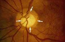
Glaucoma is an eye disease in which pressure inside the eye (intraocular pressure) rises dangerously high, damaging the optic nerve and causing vision loss. In a healthy eye, fluid is produced in the ciliary body, enters the eye, and then drains through tiny passages called the trabecular meshwork. In people with glaucoma, these passages become blocked and intraocular pressure rises.
Call Today to Schedule Your Consultation
Glaucoma Surgical Treatment Options
Some cases of glaucoma can be treated with medications. For others, laser or traditional surgery is required to lower eye pressure. Common surgeries include:
What Our Patients Have to Say
“Caring and professional, humorous but doesn’t let me slack off related to my own involvement in my care!” -Vitals.com
How Is Glaucoma Surgery Performed?
Dr. Maehara routinely performs the following advanced glaucoma surgeries in addition to standard trabeculectomy: Non-penetrating deep sclerectomy with Aquaflow implant, in-office SLT laser, Ahmed glaucoma drainage devices, and Canaloplasty.

All glaucoma patients are strictly monitored using:
The Humphrey® Visual Field Procedure
 The Humphrey Visual Field is a special automated procedure used to perform perimetry, a test that measures the entire area of peripheral vision that can be seen while the eye is focused on a central point.
What To Expect During The Humphrey® Visual Field Procedure
During this test, lights of varying intensities appear in different parts of the visual field while the patient’s eye is focused on a certain spot. The perception of these lights is charted and then compared to results of a healthy eye at the same age of the patient in order to determine if any damage has occurred.
The Humphrey system uses advanced blue-yellow perimetry, also known as Short Wavelength Automated Perimetry (SWAP), which is proven to detect signs of glaucoma-related vision loss earlier than other tests.
Patients with glaucoma will often undergo this test on a regular basis in order to determine how quickly the disease is progressing. The Humphrey Visual Field test can also be used to detect conditions within the optic nerve of the eye, and certain neurological conditions as well.
How Long Does a Humphrey® Visual Field Procedure Take?
This procedure is performed quickly and easily in about 15 minutes and is effective in diagnosing and monitoring the progress of glaucoma.
Optical Coherence Tomography
Optical coherence tomography (OCT) is an advanced technology used to produce cross-sectional images of the retina, the light-sensitive lining on the back of the eye where light rays focus to produce vision. These images can help with the detection and treatment of serious eye conditions such as macular holes, macular swelling, and optic nerve damage.
Patient Testimonial
“[The Most Honest Doctor Ever in the USA ] I am so glad to meet this doctor! I have been to almost 10 consultations already, Dr Maehara, is the only one told me I am not qualified for surgery (any surgery) for my eyes. He explained to me with data and graph! One of the most honest doctor I have ever seen! Thank you doc!”
Call Today to Schedule Your Consultation
How Does an OCT Imaging Test Work?
OCT uses technology that is similar to CT scans of internal organs, using a scattering of light to rapidly scan the eye to create an accurate cross-section. Unlike other imaging techniques, OCT uses light to produce high-resolution images, rather than sound or radiofrequency waves. Your doctor can evaluate and measure each layer of the retina through this image and compare it with normal, healthy images of the retina.
How Long Does The OCT ExamTake?
The OCT exam takes about 10 to 20 minutes to perform in your doctor’s office and usually requires dilation of the pupils for the best results.